- RESEARCH INFRA
- Special Theme _BIOMEDICAL
본문영역
The microscopic world seen through the eyes of ‘optics’
UNIST-Optical Biomed Imaging Center (UOBC)
Now that nanotechnology has entered the world of science, it has become important to look into the invisible microscopic world. Naturally, the role of optical equipment has become important, too. UNIST established its specialized center, which is equipped with the largest optical equipment in Korea, back in 2009, and is now securing global research competitiveness.

Korea’s largest biological imaging center
Biological imaging is a technique for visualizing biological activity inside the cells of the body. It can also be defined as the convergence of research on molecular cell biology and high-tech imaging technology. It is used in a wide range of fields from the identification of biological phenomena to the development of new drugs and stem cell research. As nanotechnology advances, new molecular imaging techniques using diverse nanomaterials are being developed.
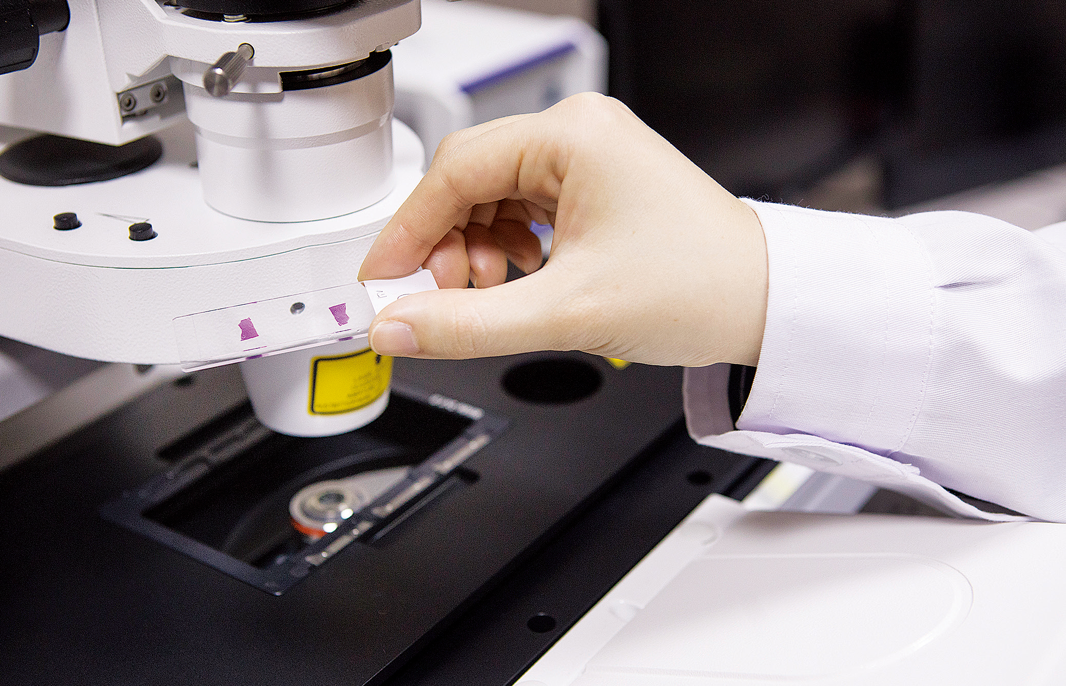
The UNIST-Optical Biomed Imaging Center (UOBC) is equipped with twenty-four types of high-tech optical equipment, which is the largest number for a single establishment anywhere in Korea. Its major items of equipment includes a ‘Super Resolution Microscope’, which allows one to observe cells with a power of up to 20㎚; a ‘Laser Scanning Confocal Microscope’, which can film the activities and movements of cells with great precision; a ‘Light Sheet Microscope’, which can film high-resolution 3D images in just a few seconds; and a ‘Deconvolution Microscope’, which features improved resolution due to the application of software functions to general optical fluorescence microscopy technology.
“In 2024, the UOBC will celebrate the 15th anniversary of its establishment. Since its inception, the center has established itself as the largest imaging center in Korea and has been assisting studies in various fields including biology, medical science, engineering, and chemistry. Recently, the UOBC has been requested for not only the biology field, but also the materials, chemistry, semiconductors, machinery, and other fields, and it also receives many external requests from other universities and research institutes for sample photography.”
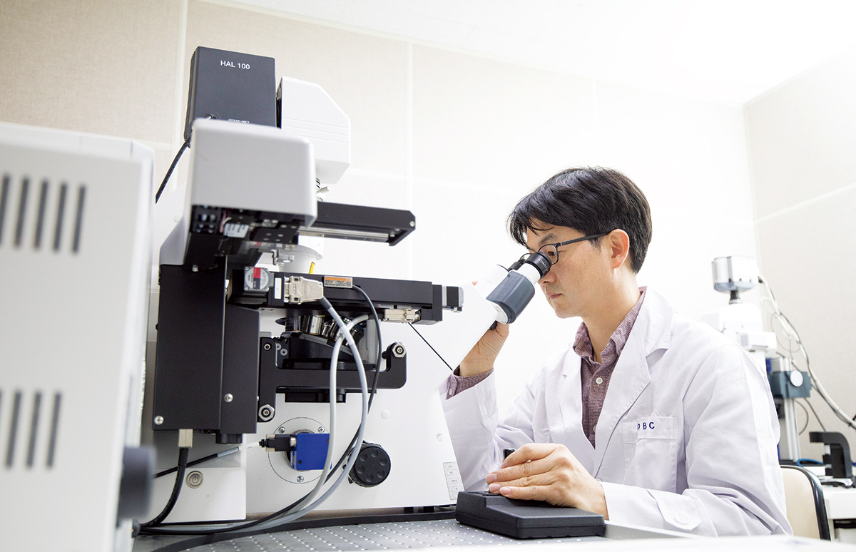
Heo Jin-hoe, a senior administrative researcher in charge of managing and analyzing the Center’s optical equipment, added that the UOBC is growing into a world-class center by continuously introducing equipment and upgrading its software. The UOBC is planning to introduce more equipment each year, and to carry out maintenance work on equipment that is over ten years old, in order to strengthen its capabilities.
UOBC, a reliable supporter of research projects
The UOBC started its business by signing an MOU with Olympus Corporation, one of the world’s top four optical brands, in October 2009. At that time, Olympus actively supported the maintenance and repair of equipment and the introduction of new equipment, and even though the MOU was terminated in 2013, they still install and operate new equipment at UNIST first, and produce better results by acquiring the latest research technology.
“The UOBC’s excellent infrastructure is a reliable supporter for UNIST’s research teams. Many centers, including UNIST’s IBS Center, Leading Research Centers, and University-centered Research Centers, are actively utilizing the UOBC’s equipment and producing great research results, and the UOBC also participates directly in some research papers.”
Recently, Professor Park Chan-young’s research team at the Department of Life Sciences carried out a study on entosis, which acts as a nonapoptotic cell death mechanism in which cancer cells can be killed, by using the UOBC’s ‘confocal laser microscope’, and a ‘single plane illumination microscope’ to accurately identify intracellular structure. Based on this work, the team studied a new mechanism of entosis and is about to publish their paper in an academic journal.
The UOBC supports not only research teams but also start-up companies founded on the UNIST campus. A representative example of this is its cooperation with ‘Load International Co., Ltd.’, which is developing fluorescent carbon particles using coffee waste. When we drink coffee, only 0.2% of the actual coffee beans are used to make a cup of coffee, while the remaining 99.8% is discarded as coffee waste. As its key business item, ‘Load International Co., Ltd.’ uses this coffee waste to synthesize carbon nanoparticles and carbon nanotube aerogel derived from coffee waste.
“The Load research team paid attention to carbon and polyphenols contained in coffee waste. While studying materials using this method, we developed ‘fluorescent carbon nanoparticles (C²NP)’. These carbon nanoparticles emit light of a specific color when they are exposed to certain wavelengths, so they are used as a key material in bio-imaging, fluorescent paints, displays, and solar cells. At the UOBC, we conducted a toxicity test on cells and a test to see whether fluorescent substances can be tinted well on living cells.”
In the future, the UOBC expects to play an important role not only in scientists’ researches, but also in product research and development for companies, once it can create an even better research environment with great ideas.
Essential infrastructure for fusion research
The UOBC is expanding its roles and areas of interest because the way to apply imaging technology has diversified greatly along with the development of the technology. In the materials and machinery fields, a lot of research has been carried out recently in connection with the bio-industry. The center is directly studying materials using bacteria or cells, or grafting them on to applied fields. It is used in diverse fields, such as biomimetic experiments, and in mechanically creating an environment that is helpful for cell culture.
“Another advantage of the UOBC is its separate operation of a sterile cell culture room. A laboratory where cell culture is difficult can use the UOBC’s internal facility and receive the center’s assistance with cell culture methods.”
According to Senior Researcher Heo Jin-hoe there is an increasing tendency to conduct convergence research in bio and other fields, so the role of the UOBC is expected to grow even more in the future. In addition, even if researchers have a good idea, there remain many difficulties in applying actual biotechnology to research, but he is convinced that the UOBC could be the answer for them.
Furthermore, researchers sometimes design together an experiment to use fluorescence at the UOBC. Although fluorescence is rarely used in research fields other than bio, the use of a fluorescence microscope allows researchers to observe and identify the structures of materials that cannot be used with an electron microscope because of the materials’ soft or moist nature. In particular, the input of fluorescence enables researchers to observe many things that couldn’t observed before, so they may be able to find clues to new research methods. With this principle, the center has supported and assisted testing in the environmental materials field using fluorescence to identify the overall structure of cement and other materials.
Such research support has been possible because the UOBC not only has high-tech optical equipment, but also employs experts who have more than ten years of experience in providing technical support to Olympus and other optical companies, including senior administrative researcher Heo Jin-hoe, as well as over ten years of experience in equipment maintenance and research assistance. As these experts design tests together and provide ideas and know-how, researchers can find a solution for imaging any special or difficult sample.
-

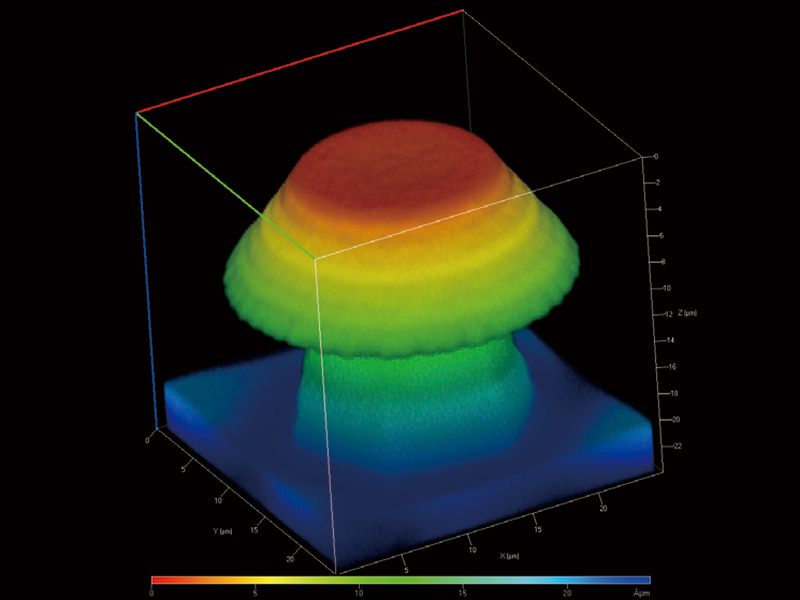
Production of a 3D Microstructure using PDMS (Polydimethylsiloxane) 3D imaging with a confocal microscope
-

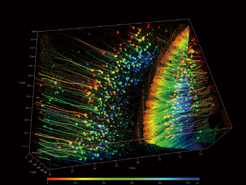
. 3D measurement of surface roughness after processing a metal surface 3D imaging with a confocal microscope
Autonomous use allowed after completion of prior training
The current method of operation supports researchers’ autonomous use of the equipment. Of course, a person who wishes to use the UOBC’s equipment autonomously needs to complete a prescribed training course. The center provides regular training programs on major equipment and associated devices, and those who finish the program and pass the exam can freely use the UOBC’s equipment 24 hours a day. As regards the use of the popular confocal microscope, many people are on the waiting list, so advance reservation is required. However, in most cases people can select and use a microscope according to their needs, so they can carry out imaging-related tests without any restrictions at the center.
“Users can directly conduct their test at any time, but if any difficulties arise, the UOBC’s professional personnel will share and discuss papers together with them in order to solve the problems while thinking about the set value for the test. We do our best to provide all possible support to provide the necessary research techniques and produce good results through the UOBC.”
Each year, the UOBC’s professional personnel participate in a number of research papers as co-authors of joint research, as they are always looking for new research techniques to apply to real studies to strengthen their expertise. We plan to continuously expand not only our joint research but also participation in papers in order to support more research fields in the future.
Senior researcher Heo Jin-hoe said that the UOBC is a place that strives to assist researchers with their study processes and results, and welcomes anyone whenever they need help. He also added that the UOBC intends to find solutions together with researchers and help them with their research even if there are limitations with the equipment.
The UOBC will celebrate its 15th anniversary in 2024. We hope that the UOBC, a great cooperative institution that drives UNIST’s research competitiveness, will grow into a world-class imaging center in the future.


